
Examining the impact of miR-379 on cancer hallmarks in an immune competent model of metastatic breast cancer
Elan C McCarthy (1), L Lin (1), K Wynne (2), S Hynes (3), CM Coleman (4), CP O’Neill (1), RM Dwyer (1,4,5)
(1) Discipline of Surgery, Lambe Institute for Translational Research, University of Galway, Galway H91 YR71, Ireland (2) Systems Biology Ireland, University College Dublin, Belfield, Dublin 4, Ireland (3) Discipline of Pathology, Lambe Institute for Translational Research, University of Galway, Galway H91 YR71, Ireland (4) Regenerative Medicine Institute, University of Galway, Galway H91 W2TY, Ireland (5) CÚRAM, SFI Research Centre for Medical Devices, University of Galway, Galway H91 W2TY, Ireland

Introduction
MicroRNA-379 (miR-379) has been reported by our group and others as a potent tumour suppressor miR with data from In Vitro, In Vivo, and clinical samples supporting potential for a compelling therapeutic function in breast cancer. This study aimed to further investigate the mechanism of action of miR-379 and a potential role in immune modulation in an immunocompetent model of metastatic breast cancer.
Materials and Methods
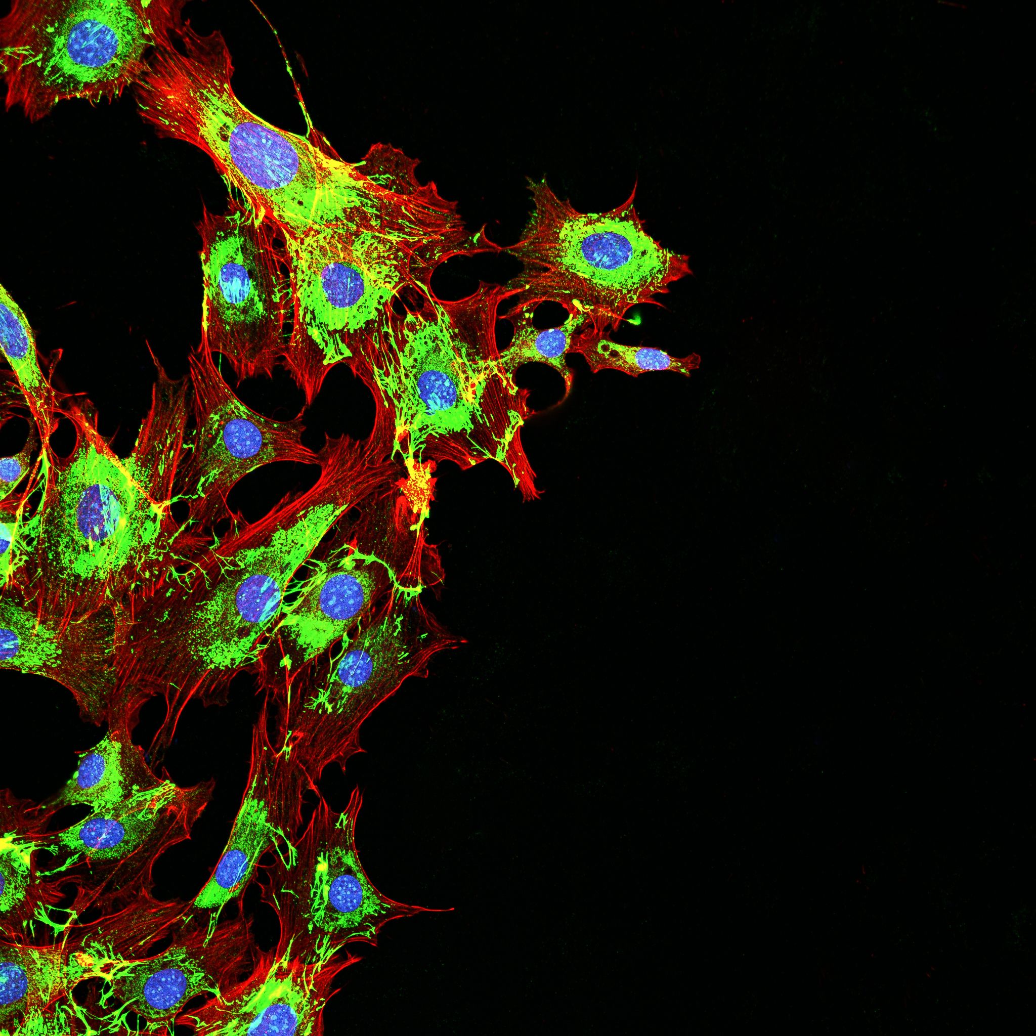
4T1 murine breast cancer cells expressing luciferase (4T1) were transduced with miR-379 (4T1-379). Balb/c immunocompetent mice received an orthotopic injection of 4T1 (n=8) or 4T1-379 (n=8) cells with n=8 mice receiving no cells. Bioluminescent (IVIS) imaging was employed to monitor disease progression. At study endpoint, tumour, lung, and bone tissues were harvested and fixed in RNAlater or 4% paraformaldehyde for proteomic or immunohistochemical (IHC) analysis respectively. Mass spectrometry was performed to determine proteins differentially regulated by miR-379. Tissue sections were probed for selected hallmarks of cancer including proliferation (Ki67), angiogenesis (CD31) and immune response (CD80, CD206).
Results and Discussion
IVIS imaging reflected tumour progression with a leading edge and subsequent metastasis to distant organs. This was reinforced by Haematoxylin and Eosin staining revealing infiltration of tumour cells into lung tissue and significant remodelling of bone evident in tumour bearing animals compared to healthy controls. A significant reduction in Ki67 was found in the 4T1-379 tumours compared to 4T1 samples (P=0.01) quantified through QuPath. Reduced expression of CD31, a marker of neo angiogenesis was also observed. Proteomic analysis revealed dysregulation of proteins associated with angiogenesis, immune response, and wound healing in tumours with elevated miR-379 expression. Furthermore, proteins associated with alternative M2 macrophage activation (Arginase-1 and Siglec1) were found to be significantly downregulated in miR-379 enriched tumours. Preliminary analysis using IHC supported this finding with an apparent increase in M1 macrophage associated CD80 and corresponding decrease in M2 macrophage associated CD206. This highlights a potential role for this miR in macrophage polarisation.
Conclusion: The immune competent model employed recapitulates Stage IV breast cancer with evidence of distant metastasis. The promising data presented highlights a potential role for miR-379 in regulation of tumour cell proliferation and modulation of immune response to cancer, which may have important implications for patient response to therapy.